


Osteochondral defects (OCD) involve damage to both the articular cartilage and the underlying subchondral bone. They occur most commonly in the knee (medial femoral condyle) and ankle (talar dome) after acute trauma, repetitive stress, or avascular necrosis of the subchondral bone. The defect leaves a region of the joint surface without the normal load-bearing and shock-absorbing properties of healthy cartilage.
Load management is the most critical variable in OCD management regardless of whether treatment is conservative or surgical. A walking cane reduces the repetitive load through the defect site with each step, which is directly relevant to reducing pain and protecting the repair or graft site.
Load at the Osteochondral Defect
In the knee, focal cartilage defects bear concentrated load during the specific knee flexion angle where the defect contacts the opposing surface. This angle varies by defect location. The total joint load reduction from a cane (15-25% per step) is distributed across the joint surface -- the proportional reduction at the defect site depends on contact angle and activity. For most OCD patients who walk with normal flexion angles, the cane reduces the total load that passes through the defect site.
Conservative Management
Small OCD lesions in skeletally immature patients (where the growth plate is still open) often heal with protected weight-bearing over months. In adults, conservative management is less effective for significant lesions but is tried for small, stable defects. Protected weight-bearing protocols (sometimes toe-touch or partial weight-bearing) may be specified -- in these cases, crutches rather than a single cane provide the weight-bearing restriction level required.
Post-Surgical OCD Repair
Several surgical approaches exist: microfracture (stimulates fibrocartilage repair), OATS (osteochondral autograft transfer), ACI/MACI (autologous chondrocyte implantation), and OCA (osteochondral allograft). Each has different post-operative weight-bearing requirements:
| Procedure | Weight-Bearing Protocol (Typical) | Single Cane Phase |
|---|---|---|
| Microfracture | NWB/toe-touch 6-8 weeks, then progressive | Weeks 8-12 (after crutch phase) |
| OATS | NWB/toe-touch 4-6 weeks, then progressive | Weeks 6-10 |
| ACI/MACI | Partial WB 6 weeks, progressive over months | Weeks 6-12 (per protocol) |
| OCA (allograft) | NWB 6 weeks, very gradual progression | Weeks 10-16 (per protocol) |
These timelines are approximate and always superseded by the surgical team protocol. OCD repair is among the longer rehabilitation protocols in orthopaedic surgery -- total recovery to sports activity may be 12-24 months.
Ankle OCD Specific Considerations
Talar dome OCD is less common than knee OCD but creates significant walking limitation. Post-operative management typically involves a boot or cast followed by progressive weight-bearing with a cane. Ankle OCD tip selection is important: the Steady Tip provides slip security on the variable surfaces encountered during the prolonged walking-return phase.
Height During Extended Recovery
OCD rehabilitation is extended (months, not weeks). The same cane may be used at different weight-bearing levels over a 6-12 month period. The collet mechanism allows precise height adjustment as gait normalises and body position changes. A single quality cane through the full recovery period is more practical than multiple canes for different phases.
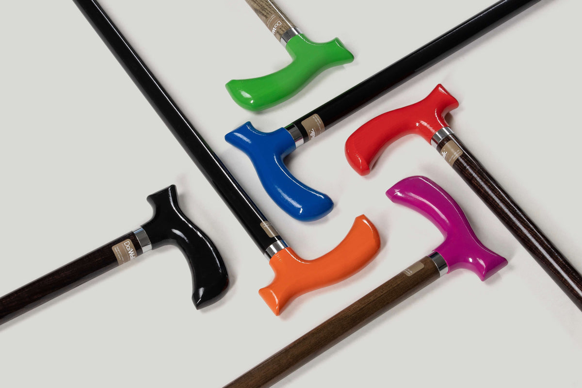
View the DaiWalk range for extended recovery cane options with the collet mechanism for sustained precise adjustment.


